
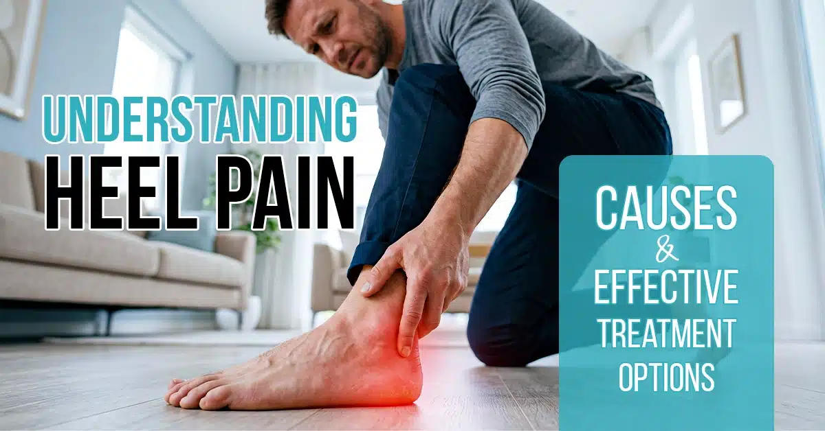
Heel pain is one of the most common musculoskeletal complaints seen at physiotherapy clinics across Ontario—and one of the most frequently mismanaged. Patients are often told to rest, ice, and stretch. Some are prescribed orthotics or sent for imaging before any clinical assessment of the tissue causing the pain has been completed. Many improve partially, then plateau, then resign themselves to managing an ongoing problem that should have resolved within three to four months of proper treatment.
At Physio Village, heel pain assessment begins with the hands—palpation, provocation testing, and movement analysis—not a scan. Identifying the precise structure at fault determines whether the treatment should be hands-on soft tissue release, joint mobilisation, nerve mobilisation, or a combination of all three. That precision is what separates full recovery from perpetual management.
What Is Heel Pain? Understanding the Anatomy
The heel is the load-bearing anchor of the foot. During normal walking, it absorbs and disperses impact forces equivalent to approximately 1.2 times body weight with every step. During running, that load increases to 2.5 to 3 times body weight. The structures most commonly injured at the heel are:
- The plantar fascia — a thick band of fibrous connective tissue running from the calcaneus (heel bone) to the base of the toes. It supports the arch of the foot and absorbs tensile load during the push-off phase of walking.
- The Achilles tendon — the largest tendon in the body, attaching the gastrocnemius and soleus muscles to the posterior calcaneal surface. It transmits the entire propulsive force of the calf during walking and running.
- The retrocalcaneal bursa — a small fluid-filled sac between the Achilles tendon and the calcaneus, prone to inflammation when the tendon is compressed during dorsiflexion.
- The calcaneal fat pad — a specialised adipose structure under the heel designed to absorb impact. Atrophy of this pad—common with ageing and corticosteroid injection—is an underappreciated foot pain cause.
Understanding which of these structures is involved determines the entire treatment approach. Treating plantar fasciitis pain with protocols designed for Achilles tendinitis, or vice versa, is not just ineffective—it can worsen the injury.
Plantar Fasciitis — The Primary Cause of Heel Pain
Plantar fasciitis pain is the most common foot complaint presenting to physiotherapy clinics in Ontario, accounting for roughly 80% of heel pain cases. It is characterised by sharp, stabbing pain at the anteromedial heel—the point where the plantar fascia inserts into the calcaneus—that is worst with the first steps in the morning or after prolonged sitting, and that typically eases after 10 to 15 minutes of walking before returning with extended activity.
Despite its name, plantar fasciitis is not primarily an inflammatory condition. Histological studies consistently demonstrate degenerative changes—collagen disorganisation, fibroblast infiltration, and neovascularisation—rather than acute inflammatory cell infiltration. This distinction matters clinically: treating it with anti-inflammatory approaches alone (ice, NSAIDs, corticosteroid injections) addresses a pathology that is not primarily present, while doing nothing to restore the tissue quality and mechanical load distribution that drive the condition.
What Causes Plantar Fasciitis Pain?
The root mechanical cause is excessive tensile load on the plantar fascia relative to its capacity to tolerate that load. Several factors contribute:
- Reduced ankle dorsiflexion range—commonly from gastrocnemius or soleus tightness—forces the foot into a supinated position during mid-stance, increasing fascial tension
- Weakness of the intrinsic foot muscles and the peroneals reduces the dynamic arch support that offloads the plantar fascia
- Excessive subtalar pronation during loading response increases the tensile strain on the medial fascial bands
- A sudden increase in load—new running volume, prolonged standing at a new job, weight gain—that exceeds the tissue’s adaptive capacity
Each of these drivers requires a specific clinical response. A rigid orthotic may reduce pronation but does nothing for ankle dorsiflexion restriction or intrinsic foot weakness—and can delay the restoration of normal foot mechanics if used as a long-term solution rather than a temporary adjunct.
Achilles Tendinitis — The Second Most Common Heel Pain Cause
Achilles tendinitis is an overload tendinopathy affecting the portion of the Achilles tendon either at its insertion into the calcaneus (insertional Achilles tendinopathy) or approximately 2 to 6 centimetres above it (mid-portion Achilles tendinopathy). The distinction between these two presentations is clinically important: they have different mechanical drivers and respond to different treatment protocols.
Mid-portion Achilles tendinopathy responds well to progressive tendon loading—carefully prescribed eccentric and heavy slow resistance exercise builds tendon capacity over 12 weeks. But this loading must be preceded by manual assessment of the calf complex, ankle joint mechanics, and tibial nerve mobility, as restrictions in any of these structures amplify the load on the tendon.
Insertional Achilles tendinopathy is more complex. The insertion is a compression-sensitive zone—the tendon wraps around the posterior calcaneus during dorsiflexion, and excessive dorsiflexion loading provokes the insertional fibres directly. Eccentric exercises that drive the heel below the step—a standard protocol for mid-portion disease—are contraindicated here. Treatment is heavy slow resistance exercise in a pain-free range, combined with manual therapy to address the ankle mechanics and calcaneal position that drive calcaneal compression.
Confusing these two presentations—or applying a generic “Achilles stretching” programme to either—consistently prolongs recovery.
Heel Spur Pain — Is It the Same as Plantar Fasciitis?
Heel spur pain and plantar fasciitis pain are commonly confused, and frequently they coexist. A calcaneal spur—a bony outgrowth from the anterior inferior surface of the heel bone—is found on imaging in approximately 50% of plantar fasciitis patients, but also in 15 to 25% of asymptomatic adults. The spur itself is rarely the direct source of pain.
What drives symptoms is the plantar fascial tension and the soft tissue irritation around the spur—not the spur itself. This is why heel spur pain responds well to the same manual therapy and loading management approaches used in plantar fasciitis, and why surgical spur removal produces disappointing outcomes in the absence of structural nerve or tendon pathology.
Manual Therapy for Heel Pain Relief — The Physio Village Protocol
The Physio Village approach to heel pain treats both the tissue directly at fault and the mechanical chain that is loading it excessively. Addressing only the heel produces temporary relief. Addressing the heel, the ankle, the calf, and the neural tissue produces lasting recovery.
Plantar Fascia Release
Direct manual release of the plantar fascia uses sustained deep pressure, cross-fibre friction, and instrument-assisted soft tissue mobilisation to address the degenerative tissue changes within the fascia. The goal is not to break down scar tissue with brute force—it is to remodel collagen fibre alignment, stimulate the tenocyte population, and reduce the adhesive connections between the fascia and the underlying intrinsic foot musculature that restrict normal toe extension.
At Physio Village, plantar fascia release is combined with active dorsiflexion movements during treatment to load the tissue under manual tension—a technique that accelerates collagen remodelling compared to passive pressure alone.
Gastrocnemius and Soleus Release for Heel Pain Relief
Restricted ankle dorsiflexion is present in the majority of plantar fasciitis patients—and the restriction nearly always originates in the gastrocnemius-soleus complex. Posterior calf tightness increases the tensile load on the plantar fascia during every step of the gait cycle.
Soft tissue release of the gastrocnemius, soleus, and Achilles tendon paratenon reduces this restriction at source. Patients treated with calf release alongside plantar fascia work consistently demonstrate greater and faster improvements in dorsiflexion range than those treated with plantar fascia techniques alone.
Joint Mobilisation of the Foot and Ankle
Restricted subtalar, talocrural, and midfoot joint mobility are common findings in patients with chronic heel pain. These restrictions alter foot mechanics during the loading phase of gait—increasing both plantar fascial tension and tibiotalar joint compressive load.
Subtalar mobilisation—specifically posteroanterior glides applied to the calcaneus—restores talar rocking motion that is essential for shock absorption during heel strike. Talocrural anteroposterior glides restore the dorsiflexion range that cannot be achieved through calf stretching alone. Midfoot mobilisation restores the intermetatarsal and navicular-cuneiform joint play that allows the arch to deform appropriately under load.
These techniques require clinical assessment first. Mobilising in the wrong direction at a restricted joint does not produce improvement—it can worsen symptoms if the joint is already irritated. The physiotherapists at Physio Village’s Oakville and Brampton locations perform joint-specific assessment before applying any mobilisation technique.
Tibial Nerve Mobilisation
A subset of patients presenting with heel pain have a neurogenic component—specifically, irritation or restricted mobility of the tibial nerve in the tarsal tunnel. This presentation is often missed, because the pain pattern overlaps significantly with plantar fasciitis.
Clinical signs that suggest neural involvement include: pain that radiates from the heel into the arch or toes, paresthesia (tingling or numbness) in the plantar surface of the foot, and symptoms that are provoked by lumbar flexion or straight leg raise testing in addition to local palpation.
Neural mobilisation of the tibial nerve—using combined ankle dorsiflexion, toe extension, and hip movement sequencing—restores neural mobility without provoking the nerve. Patients with an undiagnosed neural component do not respond to plantar fascia and ankle techniques alone, regardless of how well those techniques are executed.
Self-Management Between Physiotherapy Sessions
Heel pain management between clinic visits reinforces the progress made in treatment and prevents the mechanical re-loading that causes symptom recurrence:
- Load management — the single most important variable. Total daily step count, surface hardness, and footwear all affect fascial load. The physiotherapist will advise on appropriate activity levels for each stage of recovery.
- Intrinsic foot strengthening — short foot exercises, toe spread and press, and single-leg calf raises (at the appropriate stage of recovery) build the dynamic support that offloads the plantar fascia during walking.
- Footwear assessment — shoes with adequate heel cushioning and arch support reduce fascial load during the acute phase. Barefoot walking on hard surfaces is contraindicated until the tissue has fully remodelled.
Taping—specifically low-dye plantar fascia taping or calcaneal anti-pronation taping—provides meaningful pain relief for many patients in the early phase of treatment. The physiotherapist will apply it in clinic and teach the patient to apply it at home for the days between sessions.
Heel Pain Treatment at Physio Village — Oakville and Brampton
Physio Village treats heel pain with a structured, three-phase protocol that begins with manual therapy to address tissue quality and joint mechanics, progresses to active loading and strengthening, and concludes with a return-to-activity programme tailored to the patient’s specific demands—whether that is returning to recreational running in Brampton or managing long days on hard floors in an Oakville workplace.
Acute plantar fasciitis presenting within six weeks of onset typically resolves in 6 to 8 sessions. Chronic cases of six months or longer generally require 10 to 16 sessions. No physician referral is required for an initial assessment.
Frequently Asked Questions — Heel Pain
What is the most common cause of heel pain?
Plantar fasciitis is the most common cause of heel pain, accounting for approximately 80% of cases in clinical practice. It produces sharp, stabbing pain at the anteromedial heel that is worst with the first steps after rest and typically eases briefly after a few minutes of walking before returning with sustained activity.
How long does heel pain take to resolve with physiotherapy?
Acute plantar fasciitis presenting within six weeks of onset typically resolves within 6 to 8 physiotherapy sessions with manual therapy and structured loading. Chronic heel pain of six months or longer generally requires 10 to 16 sessions. Achilles tendinopathy has a longer natural recovery timeline—12 weeks is standard for mid-portion disease with a properly supervised loading programme.
Does stretching cure plantar fasciitis pain?
Stretching addresses the gastrocnemius-soleus tightness that contributes to plantar fascia loading, and it is a useful adjunct to treatment. On its own, without manual therapy to address the tissue changes within the fascia, joint restrictions in the foot and ankle, and intrinsic foot weakness, stretching produces partial and temporary improvement in most cases.
Should I get an X-ray or MRI for heel pain?
Imaging is not necessary for the initial assessment and treatment of heel pain in most cases. A thorough clinical examination by a physiotherapist will identify the structure at fault and determine the appropriate treatment without waiting for imaging results. If symptoms do not respond as expected after 6 to 8 weeks of appropriate treatment, targeted imaging may be indicated to rule out stress fracture or tarsal tunnel compression.
Are orthotics necessary for heel pain treatment?
Orthotics can reduce plantar fascial load in the short term and are a useful adjunct during the acute phase of treatment. They are not a long-term solution—they do not address the ankle mobility restrictions, intrinsic foot weakness, or tissue changes that drive the condition. At Physio Village, orthotics are prescribed selectively and as part of a broader rehabilitation programme, not as a standalone intervention.
Key Takeaways — Heel Pain
- Heel pain has multiple distinct causes—plantar fasciitis, Achilles tendinitis, heel spur pain, and neural compression require different treatment approaches
- Plantar fasciitis is primarily a degenerative condition, not an inflammatory one—treatment must address tissue remodelling and mechanical load distribution
- Manual therapy to the plantar fascia, calf complex, foot and ankle joints, and tibial nerve produces faster and more complete recovery than stretching alone
- Heel spur pain and plantar fasciitis frequently coexist, but the spur itself is rarely the pain source—treating the fascial tension resolves both
- Self-management between sessions—load management, intrinsic strengthening, and appropriate footwear—is as important as clinical treatment
- Physio Village in Oakville and Brampton treats heel pain with a sequenced manual therapy and loading protocol that addresses root mechanical causes, not just symptoms
Book Your Heel Pain Assessment
Share this guide: Do you know someone in Oakville or Brampton struggling with morning heel pain that never fully resolves? Share this clinical guide to help them understand what is actually driving their symptoms.
2-Minute Check: Sit in a chair with one foot resting flat on the floor. Place your thumb firmly on the inside edge of your heel bone—approximately 2 centimetres forward from the back of the heel—and press firmly upward. If that produces sharp localised pain that mirrors your walking pain, plantar fasciitis at the fascial insertion is the likely diagnosis. Then stand and take three steps. Pain worst on the first step that gradually eases is characteristic of plantar fasciitis. Pain that is constant and does not ease with walking suggests a different structure is involved—either Achilles-related or neural. Both findings warrant a clinical assessment.
Book Your Assessment: